Sunday, April 6, 2014

Sheep Brain dissection

  1. The brain of the sheep when compared to the human brain is similar in having delineated structures such as the cerebellum, the pineal gland and thalamus, and the sheep's brain even has hemispheric lobes with ventricles. The orientation of the sheep's brain is much more elongated than for that of a human brain. Another difference, which may take a moment to fully appreciate, is that the size of the olfactory bulb is much larger in the sheep's brain. As a herd animal, sheep survival often relies on the ability to detect the scents of predators. The sheep brain is also lacking the characteristic folds and large size of the human cerebrum, which explains why, so far, sheep are not hired at call centers to resolve problems that require complex language skills and decision making.
  2. The "dura mater" is the outermost layer of the membranes or meninges that protect the brain and also encases the spinal cord. Its outer side rests against the skull itself while its inner side is next to the arachnoid meninges. The dura mater part is the slick, gray lining wrapped about the brain that is only slightly opaque and seems to act like cellophane to keep the moist brain tissues moistened with cerebrospinal fluid which serves "as a liquid shock absorber around the brain and spinal cord." [Johnson, Michael D. Human Biology Concepts and Issues. 2014. Print] 
  3.  The bumps of the cerebrum, which are known as "gyri" and the grooves, which are termed "sulci" create a larger surface area while maintaining the most compact profile possible. Accordion folding is a natural design that engineers are adopting to enable storage of greater amounts of data. Brain tissues in this region process memories, reasoning, and perform higher functions that require or derive from this anatomical design. 
  4.  Dark spider web tissues clinging to the surface structures of the sheep brain are part of the blood vessel network that provide a nourishing level of oxygen to the brain, which is vital for the functions that support life. The interior brain tissues are bathed not in blood but in cerebrospinalfluid (CSF) that circulates and can be found in the ventricles. This helps to regulate the brain in such a way that typically, but not always, keeps blood-borne pathogens and drugs from interfering with brain function.
  5.  Examining the dissection of the sheep brain allows us to compare and think about the role of the pineal gland. In the sheep, like the human, this endocrine gland serves to regulate sleep patterns.Melatonin is a hormone that this gland can make or secrete so that humans and mammals get sleepy and can then perform a number of rejuvenating tasks during sleep. People who work the graveyard shifts are fighting their pineal gland's native intelligence that guides us for rest at night and alertness during the daytime hours. The pineal gland works in concert with other endocrine glands to regulate sex drive and other important functions. Flouridation of water in the drinking water supplies is reportedly having an adverse affect on human pineal glands. Research suggests a causational link between flouridation and calcification of the pineal gland in humans, which obviously lessens functionality. While one can certainly find reasons to support flouridation and it was originally propagated as a way to reduce dental caries in children, calcification of the pineal gland is no small risk.  There are a number of indications that radiation from cell phone use is also playing a role in diminished pineal gland function and disturbed sleep patterns. While our species will evolve and adapt to environmental influences over time, individual organisms will suffer in terms of function according to their genetic or constitutional predispositions and their exposure to environmental influences.

Adeloye M.B., B.S., C.P., F.R.C.S, Adelola and Felson M.D., Benjamin, “Incidence of Normal Pineal Gland Calcification in Skull Roentgenograms of Black and White Americans” American Journal of Roentgenology November 1974. Web. 5 April 2014 http://www.ajronline.org/doi/abs/10.2214/ajr.122.3.503


     6.    Ventricles are cavities or spaces within an organ such as the brain or heart through  which fluids are circulated. 

     7.     The cerebellum structure of the brain sits like a rock outcropping above the brain stem region at the back or base of the skull. Dancers, runners, and every person who enjoys fine motor skills can thank this part of their brains for providing us with balance and abilities to exhibit graceful, fluid and rapid physical movements. Alcohol has an adverse effect on this function, and the uncoordinated and sloppy movements so characteristic of people who have had too much to drink attest to the important job the cerebellum does for us.

     8.    The Thalamus and the Hypothalamus each perform important functions. They are neighbors in the center of the brain; the thalamus is larger and receives sensory information that it relays to the cerebrum. The hypothalamus actually is located under the thalamus and similarly monitors sensory signals such as what we see, smell, taste or hear. If our bodies feel cool or warm, the hypothalamus is going to be involved in modulating for homeostasis. When we recognize a hunger pang or sensation of being thirsty, the hypothalamus has been active.

     9.   The spinal cord, which is protected by the spinal column of bone vertebrae discs, has nerve tracts or bundles of axons on its outer borders. The mylenated wrapping for this is called white matter. Western science has alternately termed the non-mylenated structures of the spinal cord ( the neurons and neuroglial cells near the center of the spinal cord) as gray matter.  


   10.     I think I am very much surprised that my revulsion for anatomical dissection images has quietly abated. My revulsion for the sensory impressions of formaldyhyde however, is still strong and I do appreciate being able to observe and learn about brain structures without that assault on my senses.


Saturday, April 5, 2014

Histology Dissection Lab


 Nervous System Histology and Dissection Lab

Peripheral Nerve Histology (Microanatomy)


The differential staining in Slide 1 shows the different layers in peripheral nerve. Connective tissue stains blue. Outside the nerve, loose connective tissue combined with adipose cells is called epineurium. The next layer of connective tissue, denser and ensheathing different bundles of nerve fibers is called perineurium (dark Purplish-­blue in the slide). Delicate connective tissue called endoneurium surrounds each nerve fiber and appears as delicate blue fibers. The nerve fiber itself stains pink and is seen as a cylinder in the center of myelin with a lighter pink stain. The nerve fiber is labeled A (for axon) and the myelin is labeled M. A Node of Ranvier (R) is also visible.

1. Western science developed using Latin as the language root for terminology to create classification systems. Using just the three terms in bold in the paragraph above, we see a prefix of epi-, peri-, and endo- used to describe the structural elements in the slide of a nerve fiber. This coding system is helpful and consistent to describe the intricate structures of anatomy so that scientists, healthcare professionals and students can have a common naming convention and simplify complexities by establishing or seeing patterns. In this slide, the "epineurium" denotes the uppermost or outermost layer resting "upon" the other layers; the "perineurium" term--which means around--is used to describe the tissue that surrounds each nerve fiber, and "endoneurium," which uses the Latin prefix of "endo" means "within" or "inside," so we can clearly learn that something labeled as "endoneurium" is referring to the interior portion of a structure. 

2. Looking at the way that nervous tissue and muscle tissue is bundled, we can say that muscle fibers are ensheathed into fascicles like the strands of twine may be bound to create a rope or cable--except the fibers are sheathed without the braiding quality we can see in ropes and cables. One can also imagine that each fascicle contains strands of muscle fibers similar to the box of spaghetti pasta noodles. The fascicles provide a structural support that protects from injury and actually contributes to the movement process.

Nerve tissues are bundled, though their structure differs considerably from muscle tissues. Nerve tissues or "axons" are bundled in a myelin sheath that enables swift relay of electrical impulses to muscle tissues. Acting like an insulator, bio-electrical impulses can travel efficiently with this shield, thus saving the body's energy by not needing the active transport process (ATP) to do their work. 

3. The dark blue and purplish ares on the slide are the perineurium or the connective tissue that is around the nerve fibers.


 A. Node of Ranvier, indicated on the slide above by white gaps in intervals in between Schwann cells along an axon; these are unmyelinated spaces, that is, not covered by the insulating sheath., where the axon is exposed.
  B. Axon- represented by the deeper mauve pink with an elongated smooth appearance are the section of the neurons which carry electrical impulses, or bio-electric messages to the terminals where they inform muscles cells.
  C. Myelin Sheath is represented by the textured pink boundaries to either side of the axons described above. This sheath is generated by the Schwann cells along the axon.
  D. Endoneurium is the structure that is represented by blue fibers in the diagram above. The  endoneurium is the loosely connective tissue matrix that holds the neurons and blood vessels in formation inside the myelin sheath.  

Slide 3 Question:
To diagnose a nerve disease, what could the lipids tell you?

The lipids stained by the dye indicate health of the myelin sheath that can be damaged. If  a view of these lipids show deformities, gaps where the insulating blanket doesn't quite cover the neurons, this could indicate onset of multiple sclerosis.


Fasicles shown in diagram to left are like the casing on a sausage, holding the neurons in packages.

Perineurium represents the white boundary zones which have a denser quality that the interior tissues.

Endoneurium, which refers to what is inside, is what surrounds the neurons-the loose matrix located inside the fasicles.

Axons are the superhighway lane that is labeled on the above diagram.

Myelin Sheath, labeled above, is again what protective barrier the Schwann cells produce to increase the speed of electrical impulses.

Wednesday, March 12, 2014

Virtual Pathology Lab

Data Table

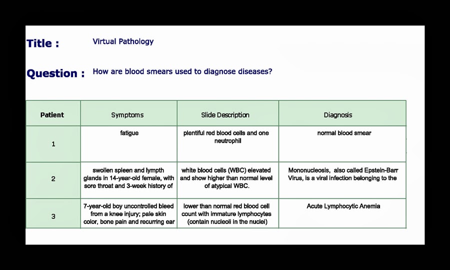
Below is a captured screen shot of the Virtual Pathology exercise case for Mononucleosis


Journal Questions 


1--In what ways do normal red and white blood cells differ?

When comparing red and white blood cells, we can differentiate based on several characteristics, such as: quantity, function, composition, and origin from the stem cells.

·        QUANTITY
Looking first at the quantities found in a person having a normal blood count, the red far outnumber the white blood cells. White blood cells (WBC) together with platelets comprise just one (1) percent of a given volume of whole blood compared to a 44 percent level of red blood cells (RBC).

·        FUNCTION
Under the right conditions, RBC are the’ worker ants of the blood’ that contain millions of proteins that tirelessly transport oxygen. In fact, a single red blood cell “can carry up to 1.2 billion molecules of oxygen.” [Human Biology textbook, p. 145] To a far lesser degree, the RBC in the blood transport carbon dioxide from the tissues.

WBC are designed to fight infection and protect the body when injured. These are the warriors of the bloodstream, the samurai who defend against invaders and have a short lifespan. They sense a threat and emit chemicals that create more WBC, calling in reinforcements when needed. Sometimes the new WBC come from the storehouse of the spleen and sometimes they are generated from the bone marrow. The WBC basically scour the body and “dine” on fungi, bacteria and parasites.

There are two types of WBC: granular and agranular. Neutrophils are the largest percentage of granular WBC and are the first responders to the scene. Other granular WBC called Eosinophils are able to act like the Lilliputians in Gulliver’s Travels who swarm and secure a larger giant. In this case, rather than stake to the ground, they use enzymes to digest unwelcome parasites. They also are responsible for chemical reactions that modulate allergic reactions in the body. A third type of granulocyte is known as the Basophils, which release histamine and help to bathe injured tissue in plasma released from nearby blood vessels.  


·        COMPOSITION
WBC are larger than RBC and are translucent-primarily because they lack hemoglobin and contain nuclei; red blood cells do not have a nucleus.


·        ORIGIN FROM STEM CELLS
Mature red blood cells, also called “erythrocytes,” derive from erythroblasts created from stem cells.  The clever design that cast out the nucleus gives the mature RBC a unique dimpled shape that makes it flexible and able to squeeze into tiny blood vessels.

All of the granular leukocytes (WBC) such as neutrophils, eosinophils and basophils, originate from stem cells by way of “myeloblasts”.

There are two agranular leukocytes (WBC), with the Monocyte originating from the Monoblasts, and the Lymphocytes originating from the Lymphoblasts.  


2. Which type of white blood cell would you expect to be most common in a normal blood smear?

Neutrophils are most commonly found in a normal blood smear


3. A differential count of white blood cells from a patient gave the absolute number of lymphocytes as 8000 per mm3 and the total number of white blood cells as 12,000 per mm3. Calculate the percentage of lymphocytes in this sample of white blood cells. Is this a normal or abnormal percentage? Explain your answer.


A normal range of lymphocytes is the 20 to 40 percentile, so this an example of an abnormally high percentage, in the neighborhood of 67 percent.  


4. Describe the difference between a communicable disease and an inherited disease. Use examples you have studied in this exploration to support your description.


Essentially, the difference between a communicable disease and an inherited disease is that you can pass a communicable disease to others primarily through vectors such as parasites, viruses, or bacteria via physical contact; transmission often occurs through exposure to blood, saliva, fluid from the mucous membranes of the lungs and airways, and other body fluids. 

Inherited diseases are of course transmitted via the genetic roulette of procreation, but having a genetic variation that fits the definition of a disease is dependent on the cellular matrix unique to offspring and mutations in genetic code that persist generation to generation. 


For example, research shows that people with chronic myelogenous leukemia, which is one of those rare diseases characterized by the bone marrow factories being overrun by abnormal and inefficient white blood cells, have an abnormal chromosome as the causal factor. However, the boundary zone for leukemia origin between communicable and inherited is fuzzy; environmental exposures and even viral infections are thought to be the causal factors in some cases.


The person cannot maintain the homeostasis and intricate symphony of balances that normally keep white blood cell production in the proportion needed for good health and a strong immune system.


5. Why are white blood cells in a stained blood smear usually counted at low power under a microscope? Explain your answer.


White blood cells are large elements in a given blood sample so it would be like taking a close up photo of a giraffe and only seeing a spot on the neck if you use the high power. Use the low power magnification when examining a stained blood smear of white blood cells and you can see and count their numbers much like a camera with a wider lens will be able to capture not just the spot, but the entire neck of the giraffe as well as the head, body and legs.


6. Why is the presence of a larger than normal number of neutrophils indicative of an infection? Explain your answer.

Neutrophils are the first line of defense and as such, they aggressively outnumber and basically engulf a foreign body or invader such as bacteria or virus before it can spend much time or do much damage. When they mass, this is a sure sign of an infection, of the immune system working as designed to thwart what does not belong.


7.  Why would you not expect to see tissue macrophages in a blood smear? Explain your answer.


Macrophages are a type of large white blood cell that are like the trash trucks that come by to remove the red blood cells that reached their expiration date of 120 days. The liver and spleen are where old RBC get destroyed. Much like the neutrophils engulf foreign bacteria or viruses, the macrophages engulf the no longer useful RBC and digest them. It’s a process called “phagocytosis” and it’s a housekeeping duty our bodies perform at the cellular level.

Blood smears are a snapshot of what is going on in the bloodstream. Since the old red blood cells are more or less sequestered in the liver and spleen for this housekeeping, the macrophages typically won’t be visible in a blood smear.  

Tuesday, January 28, 2014

1.28.2014 FORWARD

GRANULAR VIEWS

It is late, but I am getting familiar with blogging tools and experimenting in the spirit of the course. I look forward to viewing the virtual microscope that our instructor mentioned in her most recent video. I enjoy reading and learning so I have become attached to the name of Nidaba, which according to online references, has something to do with a Sumerian goddess of writing, learning and the harvest.

I will want to learn more about that. I have always been attracted to the pleasures of writing and yet, oddly enough, this is my first blog--as a student. Finding an online biology course this semester was looking like a dim prospect right up until I managed to spot one seat left and dared to inquire by email if I might be a late registrant.

Once I determine the option of keeping this blog from going viral, I will post additional comments for classmates. I am certainly not interested in following comments from anyone in the virtual world led to this blog by bots and the practice of indexing for search engines.